
Scientists just found a surprising new job for the brain's fear center, and it has nothing to do with being scared.
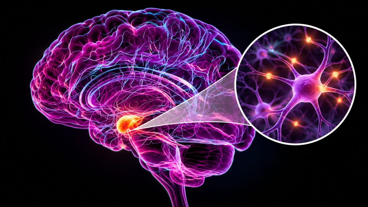
Researchers reported October 27 in Nature Neuroscience that individual amygdala memory neurons may act like a save button, flagging which everyday images a person will later remember. The study, covered by Science News on November 12, 2026, used patients with implanted brain electrodes to track single neurons in real time.
Brain-Computer Interface Speech Restoration Hits 95 Percent Accuracy in New Tests
How Amygdala Memory Neurons Were Discovered
The amygdala sits deep in the brain and has long been known as the command center for fear and aggression. For decades, scientists mostly studied it through scalp electrodes or by watching thousands of neurons fire together. That approach never showed how single cells behave during memory tasks.
This time, researchers got a rare chance to look closer. They worked with patients who already had electrodes placed deep in their brains for unrelated medical monitoring. That setup let the team record from individual amygdala memory neurons, something nearly impossible to do with typical brain scans because the structure is small and buried far beneath the skull.
Why Earwax Deters Insects: The Sticky, Slippery Trick That Stops Ants Cold
Testing Memory With Everyday Images
Participants looked at pictures of ordinary things like food and animals, sorted into five categories. After a short break of roughly 10 to 30 minutes, they were asked which images they recognized from before. Researchers then lined up each person's brain activity with their actual memory performance.
Some amygdala neurons lit up strongly for specific categories, almost like they had favorite subjects. When those category-tuned neurons fired harder during the first viewing, people were more likely to recall that exact image later. Weaker neuron responses lined up with images people simply forgot.
Researcher Xiao, quoted by Science News, said a remembered image appeared to be “earmarked for longer term storage.” Xiao also noted the study does not pin down the exact biological mechanism behind memory formation, but said the results point toward a bigger role for the amygdala in sorting what the brain keeps and what it tosses aside.
Comparing the Amygdala and Hippocampus
The hippocampus has always been the brain's go-to region for forming and retrieving memories. So researchers compared it directly with the amygdala in this same experiment. According to the report, hippocampal activity showed a weaker link to recall, and it also seemed to kick in slightly later than the amygdala signal.
That timing gap matters. It hints that amygdala memory neurons might make an early call on whether something is worth remembering, before the hippocampus even gets fully involved. Scientists are careful to note this is a proposed sequence, not a confirmed one. The two regions likely work as partners rather than rivals, each handling a different piece of the memory puzzle.
This also reshapes the amygdala's reputation. People usually associate it only with fear, panic or aggression. But this experiment used calm, neutral pictures of food and animals, nothing threatening at all. Yet the neurons still responded and still predicted memory outcomes, suggesting the amygdala's job is broader than scientists assumed.
Why the Findings Matter for Memory Disorders
Xiao pointed out that memory disorders remain a massive medical problem with no broadly effective treatments available today. Understanding how the brain decides what to keep could eventually open doors for new therapies, though that outcome is still far off.
The current study only tested short delays of half an hour at most. It says nothing yet about memories that last for days, years or a lifetime. Researchers were also careful to stress that this work shows a correlation, not proof that these neurons directly cause memories to stick. Whether manipulating them could boost or restore memory is still an open question.
What Comes Next for Amygdala Research
Scientists now want to map out exactly how amygdala memory neurons talk to the hippocampus and other memory regions. They also plan to test whether the same patterns hold up over much longer stretches of time, not just minutes.
Another big question is emotion. Since the amygdala is famous for processing fear, researchers want to know if emotionally charged images produce even stronger save signals than neutral ones like fruit or animals. Larger studies across more patients will be needed before anyone can talk seriously about turning this into a treatment. For now, the discovery simply adds a new layer to a brain region people thought they already understood.